

This year's Olympus digital photography competition , BioScapes, rewards the "dive" into the microcosm. The first place was taken by the video of American researchers that refers to a "fluffy" mass and reveals the magical process of embryonic development of a fruit fly. Photo: William Lemon, Fernando Amat, Philipp Keller/ Olympus BioScapes Digital Imaging Competition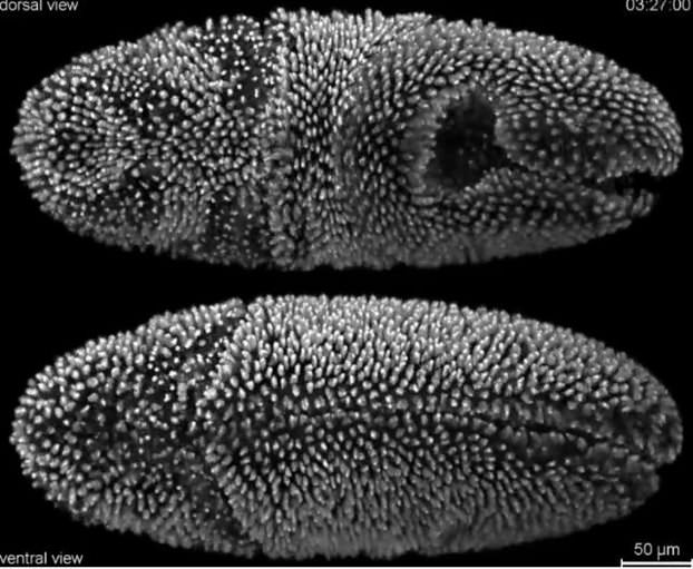
From the mysteries of life to the magic of nanoscale organisms, photography combined with microscopy has opened a new chapter in precision research. A few days ago, Olympus' digital photography competition, BioScapes 2014, awarded photographers who dared to "dive" into the microcosm, revealing the grandeur of life.
This year, the first prize was won by an impressive video about the embryonic development of a fruit fly (Drosophila, also called a fruit fly) by researchers William Lemon, Fernando Amat and Philip Keller of the Howard Hughes Medical Institute (HHMI), in Virginia, USA.
The material (which interested parties can find here: www.olympusbioscapes.com/gallery/year/2014) stood out among 2,500 entries and, in addition to first place, awarded the winners Olympus photographic equipment worth $5,000 – that is, approximately 4,100 euros.
The high-speed video, according to the Japanese giant, offers scientists for the first time the ability to monitor the "fate" of individual embryonic cells from shortly after fertilization until the larva hatches. The footage shows the process of cell division and migration, the formation of internal organs and the structure of the fruit fly's body.
A better understanding of the processes that lead to the complete formation of an organism could help experts investigate in depth the mechanisms behind the occurrence of serious diseases.
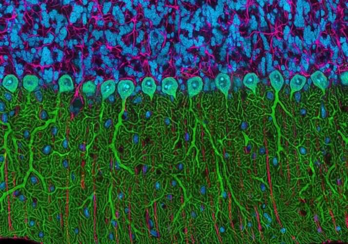
In second place was the photo of a rat cerebellum belonging to Dr. Thomas Drierink from the University of California, San Diego, specifically from the National Center for Microscopy and Imaging Research.
